Mosaicism
With the advent of new technologies for PGT-A, such as aCGH and NGS, it has become possible to detect the number of each chromosome copies. Thanks to this, we can now detect embryonic mosaicism. Embryonic mosaicism is a phenomenon characterized by the presence of two or more genetically different cell lines (cell clones), usually one with a chromosomal abnormality and one with a normal chromosomal set.
All cells in an embryo originate from a single fertilized zygote cell, but not all of them may have the same set of chromosomes later in life. Differences in chromosome set among cells are due to mitotic errors (Fig. 1),
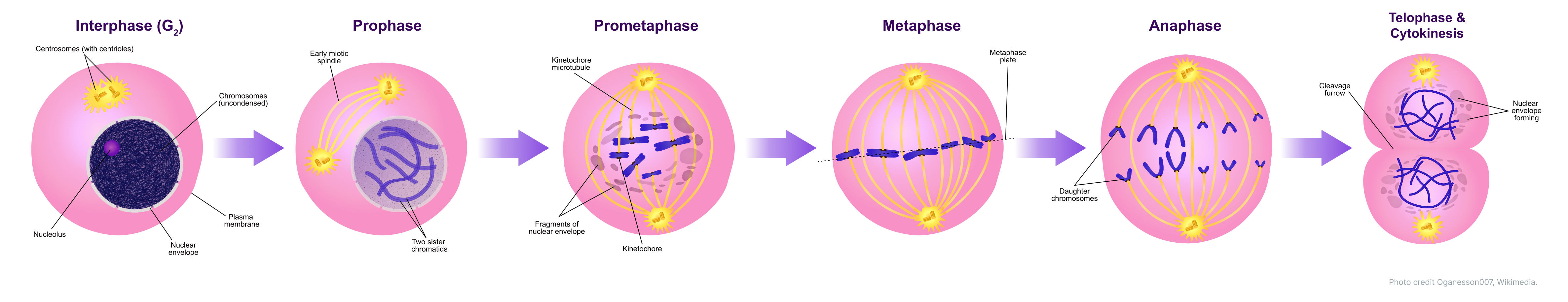
that can occur during embryonic development. Depending on which stage of postzygotic development these errors occurred, mosaicism can affect different parts of the blastocyst.
For the PGT-A test, a biopsy of 5-10 cells of the trophectoderm at the blastocyst stage is performed, while the intracellular mass (ICM) remains intact. In some cases of embryonic mosaicism, errors can occur during the evaluation of a particular biopsy. For example, when biopsying an embryo with mosaicism in the ICM cells, the trophectoderm cells will always be different, leading to an incorrect result. When biopsying an embryo with ICM/TE mosaicism, the result will only reflect the status of the trophectoderm (euploid/aneuploid) when the ICM will contain the opposite cell clone (aneuploid/euploid).
Even in embryos with mosaic trophectoderm, the detection of mosaicism will depend on the biopsy location according to the different distribution of euploid and aneuploid cells. Therefore, when detecting mosaicism in trophectoderm cells, the percentage of mosaicism in a particular biopsy cannot be extrapolated to the entire embryo. Consequently, the result obtained in a particular biopsy should be considered as referring only to the biopsy itself.
Several types of cell mosaicism have been described at the blastocyst stage (Fig. 2):
1. Complete mosaicism — mosaicism is present in the intracellular mass and in the cells of the trophectoderm.
2. ICM mosaicism — mosaicism is present only in the intracellular mass. Such mosaicism cannot be detected.
3. Trophectoderm mosaicism (TE) — mosaicism is present only in trophectoderm cells.
4. ICM/TE mosaicism (type 1) — trophectoderm cells are euploid, while ICM cells are aneuploid. Such mosaicism cannot be detected.
5. ICM/TE mosaicism (type 2) — ICM cells are euploid, and trophectoderm cells are aneuploid. Such mosaicism cannot be detected.

Scientific studies show that when several biopsies of the same embryo are performed, the level of concordance mosaicism reaches 95-100 % (Johnson DS, Cinnioglu C, Ross R, Filby A, Gemelos G, Hill M, et al. Comprehensive analysis of karyotypic mosaicism between trophectoderm and innercell mass. Mol Hum Reprod, 2010)In addition, the researchers also analyzed the ICM of the same embryos to assess the frequency of mismatches between cell lines. TE and ICM in mosaicism differ by only 3-4% (Capalbo A, Wright G, Elliott T, Ubaldi FM, Rienzi L, Nagy ZP. FISH reanalysis of inner cell mass and trophectoderm samples of previously array-CGH screened blastocysts shows high accuracy of diagnosis and no major diagnostic impact of mosaicism at the blastocyst stage. Hum Repro, 2013). Thus, trophectoderm biopsy is considered to be a good method of diagnosing embryonic mosaicism.
NGS has made it possible to detect mosaicism more accurately in trophectoderm cells, making it possible to decide to transfer mosaic embryos in the absence of euploid embryos. It should be noted that there are few studies on the clinical outcomes after mosaic embryo transfer.
In order to assess the real frequency and possible consequences of mosaic embryo transfer, the results of PGT-A should be evaluated considering three aspects: embryo quality; number of cells analyzed; frequency of implantation, miscarriages and live birth for mosaicism on that particular chromosome.
Not long ago, a study was conducted (Spinella, F, Fiorentino, F, Biricik, A, Bono, S, Ruberti, A, Cotroneo, E, Baldi, M, Cursio, E, Minasi MG, Greco E. Extent of chromosomal mosaicism influences the clinical outcome of in vitro fertilization treatments. Fertil Steril, 2018), in which mosaic embryos were transferred to patients who could not receive euploid embryos in the ART program. A total of 77 embryo transfers were performed, of which 44 were embryos with a percentage of mosaicism below 50% and 33 were embryos with a percentage of mosaicism above 50%. The data show that almost half (48.1%) of the transfers resulted in biochemical pregnancy, 38.5% resulted in implantation, one third (30.8%) of the mosaic embryo transfers resulted in live birth.
However, when comparing the euploid and mosaic embryo groups in terms of outcomes, the study data show a higher percentage of implantation (54.6% vs 38.5%), pregnancy (46.4% vs 30.0%) and live births (46.6% vs 30.8%) when euploid embryos were transferred, as opposed to mosaic embryos, which makes sense. Interestingly, when embryos with a lower percentage of mosaicism were transferred ( 50%), which may suggest that in some cases low-level mosaicism may be caused by amplification artifacts (see "Technical features"). (Spinella, F, Fiorentino, F, Biricik, A, Bono, S, Ruberti, A, Cotroneo, E, Baldi, M, Cursio, E, Minasi MG, Greco E. Extent of chromosomal mosaicism influences the clinical outcome of in vitro fertilization treatments. Fertil Steril, 2018)
In another similar study (Victor, AR, Tyndall, JC, Brake, AJ, Lepkowsky, LT, Murphy AE, Griffin, DK, McCoy RC, Barnes, FL, Zouves, CG, Viotti, M. One hundred mosaic embryos transferred prospectively in a single clinic: exploring when and why they result in healthy pregnancies. Fertil Steril, 2019) , 100 mosaic embryos with different chromosomal abnormalities were transferred: mosaic monosomy/trisomy, segmental abnormalities, and mosaic-aneuploid embryos (2 or more chromosomes); the percentage of aneuploid cells ranged from 20 to 80%. The data showed that mosaic embryos with single segmental abnormalities performed better in terms of achieving biochemical pregnancy (57.6% vs 49.0%) and implantation (45.5% vs 38.0%) than all other groups of embryos. However, embryos with mosaic segmental abnormalities should be treated with special caution because of the higher risk of birth defects associated with live birth.
The level of mosaicism determined by PGT-A of a particular biopsy is not recommended as a decisive criterion for the priority of mosaic embryo transfer. The nature of the chromosomal abnormality plays an important role: the complete chromosome or its segment, as well as which chromosome was involved.
It should also be noted that a low or high percentage of mosaicism during PGT-A can result from a noisy profile due to various technical reasons, which can lead to false results (Preimplantation Genetic Diagnosis International Society. “PGDIS POSITION STATEMENT ON THE TRANSFER OF MOSAIC EMBRYOS IN PREIMPLANTATION GANETIC TESTING FOR ANEUPLOIDY (PGT-A).” PGDIS, 2019). Such causes can occur at the embryo trophectoderm biopsy stage: a poor-quality biopsy in which too few cells are selected with damage or partial destruction and loss of cellular DNA, which affects chromosome profiles. Also at the stage of genetic analysis: during library construction, poor quality of the original DNA (uneven amplification of the whole genome) can lead to insufficient or excessive representation of chromosomes (whole chromosome mosaicism) or chromosomal regions (segmental mosaicism). Bioinformatic analysis algorithms used in normalizing chromosome bin maps can also potentially alter profiles. Our laboratory uses an advanced bioinformatic algorithm; in addition, the laboratory tracks the percentage of mosaic embryos both among all results obtained and for specific embryologists.
Patients considering mosaic embryo transfer should undergo genetic counseling, exploring the possible risks and outcomes associated with such embryo transfer (8). It should be explained to the patient at the counseling session that if a pregnancy is achieved with a mosaic embryo transfer, confirmation of the fetal karyotype by invasive prenatal diagnosis, preferably by amniocentesis, is recommended.
The International Preimplantation Genetic Diagnostic Society (PGDIS) has published updated guidelines for professionals in 2019. Key points:
1. Professionals (reproductologists, embryologists, geneticists) should inform patients that a genetic test, in this case PGT-A, based on sampling one or a small number of embryonic cells, cannot be 100% accurate, given technical and biological factors, including chromosome mosaicism.
2. If mosaic embryos are to be transferred, patients must be made aware of any potential risks, which will be noted on the informed consent form.
3. The transfer of euploid embryos should be prioritized over the transfer of mosaic embryos.
4. If a mosaic embryo transfer is considered, the following options should be discussed with the patient:
- Initiation of the next IVF cycle with PGT-A to increase the likelihood of obtaining an euploid embryo for transfer;
- Blastocyst transfer with lower mosaicism level after appropriate consultation.
Professionals should strongly recommend that patients undergo prenatal diagnosis of the fetus and placenta of any established pregnancy after PGT-A, especially after a mosaic embryo transfer. The amniocentesis assay (starting at 14 weeks) is currently considered to be the most representative for the fetal chromosomal status testing. Of the earlier tests (starting at 10 weeks), one may also consider NIPT, which analyzes the copy number of all 24 chromosomes in the fetus – the standard 5-chromosome NIPT tests for chromosomes 21, 18, 13 X and Y may not be appropriate. Ultrasound should also be used to detect fetal abnormalities, while PAPP-A screening and Doppler ultrasonography can also be used to detect placental abnormalities.
1. Capalbo A, Wright G, Elliott T, Ubaldi FM, Rienzi L, Nagy ZP. FISH reanalysis of inner cell mass and trophectoderm samples of previously array-CGH screened blastocysts shows high accuracy of diagnosis and no major diagnostic impact of mosaicism at the blastocyst stage. Hum Reprod 2013; 28.
2. Johnson DS, Cinnioglu C, Ross R, Filby A, Gemelos G, Hill M, et al. Comprehensive analysis of karyotypic mosaicism between trophectoderm and inner cell mass. Mol Hum Reprod 2010; 16:944–9.
3. Martinez MC, Mendez C, Ferro J, Nicolas M, Serra V, Landeras J. Cytogenetic analysis of early nonviable pregnancies after assisted reproduction treatment. Fertil Steril 2010; 93:289–92.
4. Hassold T. Mosaic trisomies in human spontaneous abortions. Hum Genet 1982; 61:31–5.
5. Maria Vera-Rodriguez, Ph.D. and Carmen Rubio, Ph.D. Assessing the true incidence of mosaicism in preimplantation embryos . Fertil Steril, 2017; 107, 5, 0015-0282
6. Besser AG, Mounts EL. Counselling considerations for chromosomal mosaicism detected by preimplantation genetic screening. Reprod Biomed Online 2017; 34:369–74.
7. Francesca Spinella, Ph.D., Francesco Fiorentino, Ph.D., Anil Biricik, Ph.D., Sara Bono, Ph.D.,Alessandra Ruberti, B.Sc., Ettore Cotroneo, Ph.D., Marina Baldi, Ph.D., Elisabetta Cursio, B.Sc., Maria Giulia Minasi, B.Sc., and Ermanno Greco, M.D. Extent of chromosomal mosaicism influences the clinical outcome of in vitro fertilization treatments. Fertil Steril, 2017, 0015-0282.
8. Preimplantation Genetic Diagnosis International Society. “PGDIS POSITION STATEMENT ON THE TRANSFER OF MOSAIC EMBRYOS IN PREIMPLANTATION GANETIC TESTING FOR ANEUPLOIDY (PGT-A).” PGDIS, 27, May 2019